The AACR Annual Meeting 2026 featured six days of groundbreaking science across the continuum of cancer research. With nearly 250 clinical trials presented, including several practice-changing studies, the meeting highlighted the many leaps in patient care that are on the horizon.
Of course, advances in cancer treatment are only possible because of the decades of basic and translational research that preceded them. Propelling further progress at the bench, therefore, will require ever-advancing tools that empower even more fundamental insights into how cancer forms, how it spreads, and, ultimately, how it can be stopped.
From posters to plenaries, this year’s AACR Annual Meeting presentations were full of innovative technologies that are allowing researchers to study cancer in new ways. Here, we highlight four presentations that showcased how new research tools are leading to discoveries with treatment implications.
Shaping Up Tumors to Study Cellular Plasticity
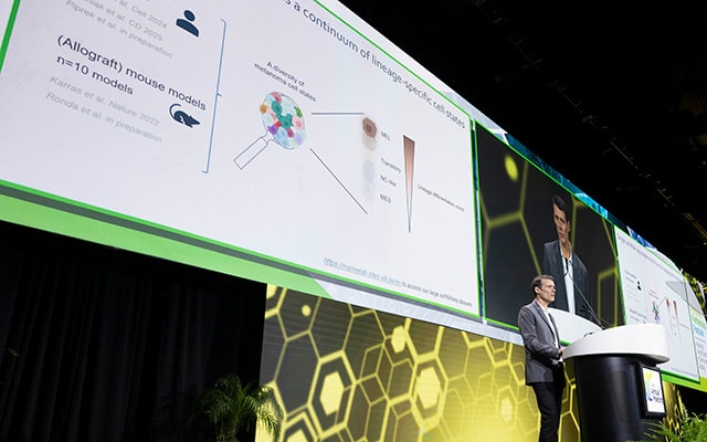
During the Discovery Science Plenary Session on minimal residual disease, Jean-Christophe Marine, PhD, of the VIB-KU Leuven Center for Cancer Biology in Belgium, shared a new physics-based framework for studying intratumoral heterogeneity and treatment-induced cellular plasticity—two features that contribute to the development of treatment resistance in melanoma.
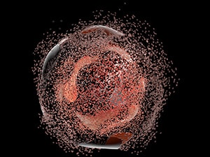
Building on observations that different cell states are associated with unique morphologies, Marine and colleagues developed ORIGAMI, which measures cell surface area and volume to define the heterogeneous cell states present in melanoma and to monitor treatment-induced transitions from one state to another that could indicate emerging treatment resistance.

“What’s interesting about this approach is that you can basically define cell state in the absence of any omic measurements,” Marine said.
Marine shared that he and colleagues applied ORIGAMI in a screen for therapeutic agents that could resensitize treatment-resistant melanoma cells, using reduction in cell surface area as a readout of treatment response. This approach revealed that treating melanoma cells with modulators of the endoplasmic reticulum and Golgi apparatus may overcome resistance to trametinib (Mekinist) and palbociclib (Ibrance) treatment. They also found that these agents could improve efficacy of immune checkpoint inhibition in cells that had developed resistance to prior targeted therapy.
Tracing Neuronal Innervation of Pancreatic Cancer
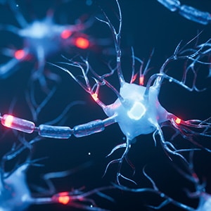
It’s long been understood that neurons infiltrate tumors, but efforts to characterize these neurons and their role in tumor biology have been hampered by the fact that the neuronal cell bodies, which house the neuron’s genetic information, are found outside of the tumor microenvironment. With only the nerve ends infiltrating tumors, the gene expression profiles of innervating neurons are not captured by conventional tumor sequencing methods.
In a Major Symposium on Neural-immune Crosstalk in Cancer, Andreas Trumpp, PhD, of Heidelberg Institute for Stem Cell Technology and Experimental Medicine in Germany, shared a technique that he and colleagues developed to overcome this obstacle.
The method, Trace-n-Seq, involves injecting a tumor with a tracer dye known as Fast Blue. The tracer is taken up by the tumor-infiltrating nerve ends and travels in retrograde on the axon back toward their neuronal cell bodies, thereby labeling neuronal cell bodies whose nerve ends are present in the tumor. After several days, the researchers harvest clusters of neuronal cell bodies and use cell sorting to isolate those positive for Fast Blue. The Fast Blue-positive neuronal cell bodies then undergo single-cell RNA sequencing.
Trumpp and colleagues applied this method in mouse models to characterize the expression profiles of thousands of neurons innervating pancreatic ductal adenocarcinoma (PDAC), whose cell bodies are typically located next to the aorta or spinal cord. They identified transcription profiles unique to PDAC-innervating neurons, as compared with neurons that innervated healthy pancreata, and observed increased axonal “sprouting” in PDAC, a branching architecture that led to more contacts between nerve ends and cancer cells.
They also used Trace-n-Seq to examine the impact of nab-paclitaxel on PDAC innervation, finding that this common pancreatic cancer treatment significantly reduced the number of innervating sensory neurons in treated tumors and led to tumor shrinkage. Further, they found that reduction of tumor innervation increased the efficacy of immune checkpoint inhibition in PDAC models. Trumpp and colleagues are now evaluating the clinical efficacy of combining nab-paclitaxel, gemcitabine, and immune checkpoint inhibition to treat PDAC.
SPARC Sparks Progress in Understanding Treatment Response
Analyzing histopathology slides to predict treatment response is a common application of artificial intelligence (AI), but current tools are usually unable to identify the mechanisms underlying response or resistance to treatment. This means researchers are missing out on key information that can reveal strategies for overcoming resistance or for treating resistant tumors.
During the Opening Plenary Session, Regina Barzilay, PhD, discussed a new AI-based model she and colleagues have developed that analyzes histopathology slides to not only predict treatment response, but also to identify the transcriptional programs associated with response.
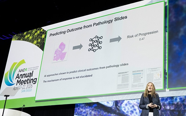
The model, called SPARC, was developed by integrating data from histopathology slides with data from spatial transcriptomics. By training on both types of data, the researchers were able to produce a model that could examine a histopathology slide and, based on the data on which it was trained, report the transcriptional programs active in different regions of the tumor tissue and predict likelihood of response.
By determining the transcriptional programs associated with treatment response or resistance, SPARC provides researchers with valuable information that can guide treatment, Barzilay noted.
“[SPARC] can give us an idea of what should be our next target for the intervention,” she said. “Using this data to identify targets is the main application of this technology.”
Tumor Acidity: Low pH, High Selectivity
Selectively targeting cancer cells while leaving most healthy cells alone is the central tenet of cancer treatment. Using targeted therapeutics designed to go after tumor-associated biomarkers is one way to achieve this, but these typically only work for patients whose tumors express the biomarker. An alternative approach that researchers are exploring is to take advantage of cancer’s acidic microenvironment.
“Tumor acidity is a universal hallmark of the microenvironment, driven by elevated glycolysis, and offers a broadly applicable opportunity for tumor-specific therapy,” said Qiang Feng, PhD, of Harold C. Simmons Comprehensive Cancer Center at The University of Texas Southwestern Medical Center, who presented his research during a Poster Session at the AACR Annual Meeting 2026 on Innovative Therapeutic Modalities and Translational Platforms.
Feng and colleagues exploited tumor acidity to more precisely deliver the immune-stimulating cytokine IL-2, which is used in cancer treatment but is associated with toxic side effects due to off-target activity.
First, they embedded ultra-pH-sensitive (UPS) nanoparticles into three-dimensional tumor cultures and human tissue to identify areas of severe acidity. The UPS nanoparticles were designed to fluoresce once the surrounding pH dropped below 5.3, allowing the researchers to map severely acidic regions. The researchers integrated these data with spatial transcriptomics and found that areas of severe acidity were enriched for cytokine-mediated signaling and cellular extravasation, both of which can indicate immune activation.
Feng and colleagues also observed that when they delivered UPS nanoparticles to patients through intravenous administration, they were activated in tumors but had minimal activation in normal organs, indicating tumor selectivity. Based on these observations, they reasoned that encapsulating IL-2 with UPS nanoparticles may enable selective delivery to tumors and avoid the side effects associated with standard administration. They evaluated the approach in mice and found that UPS-encapsulated IL-2 was just as effective as unencapsulated IL-2 in shrinking tumors but was associated with significantly less systemic interferon activation, which suggested less toxicity.
The post From the Bench, AACR Annual Meeting 2026: Innovative Tools Uncover New Insights Into Cancer appeared first on American Association for Cancer Research (AACR).